
Radiology/Imaging Department Equipment
Low cost Ultrasound Machines
Low-cost ultrasound machines are essential for providing affordable healthcare, especially in resource-limited settings. Here are some options and considerations for low-cost ultrasound machines:
Below are the Options for Low-Cost Ultrasound Machines
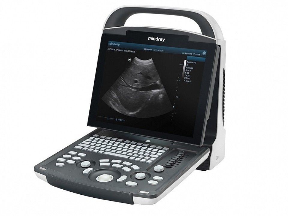
MINDRAY DP-10 WITH LINEAR PROBE
MHULDP-10
Buy Now
MINDRAY DP-10 B/W system with 1-probe connector, 12.1" high resolution LED monitor.
WED-9618 B-Ultrasound Diagnostic Apparatus

MHULWELLD-01
Buy Now
Main Unit Parameters
- Scanning Model Electronic Linear Array, electronic convex array
- Cine-loop ≥400 frames
- Image Storage ≥32 frames
- Display Depth ≥250mm
- Scanning Angle Visible and adjustable
- Display Mode B、B+B、B+M、B+M/M、M、4B
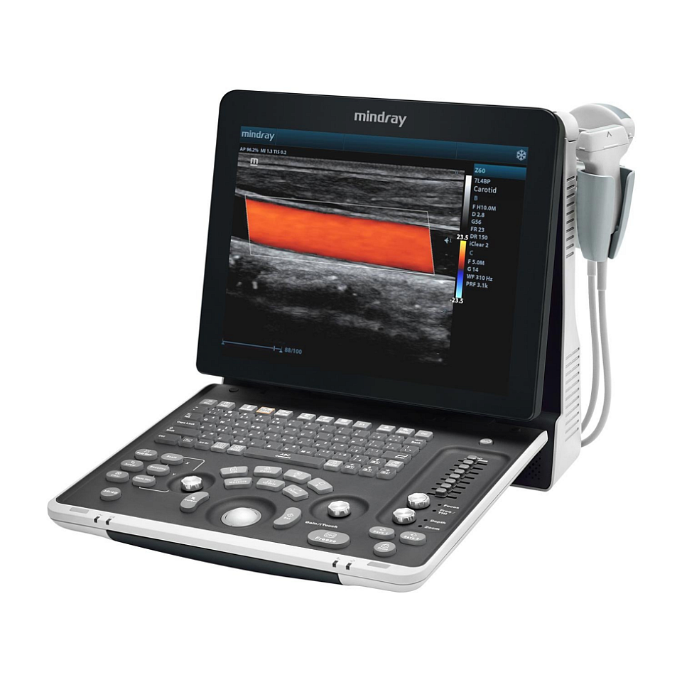
MINDRAY Z60 COLOUR ULTRASOUND with 2 probe connectors
MHULZ60-01
Buy Now
Accuracy | quality | Mobility
The Z60 is a wise choice for those who require high-quality image performance, ease of mobility, as well as affordability when it comes to advanced ultrasound imaging.
